Selling fast!
Get yours while you can.
Free Shipping on web orders over $50! (USA only. Some exclusions.) Our mid-season sale is on. Shop Phones
MICRO-EPORE
Couldn't load pickup availability
Prices valid in USA, Canada, and PR only.
The new WPI MICRO-ePORE™ pinpoint cell penetrator is a simple and versatile system that can be used for efficient cell microinjection of a diverse array of compounds and biomolecules into oocytes and pre-implantation stage mammalian embryos. Patent pending Flutter Electrode Technology assists in small, clean, precise membrane penetration without tearing or damaging the membrane when you are working on the microinjection of transgenic animals or cell manipulation.
Platinum wire electrode holders (sold separately) are available!
NOTE: Microinjection pump and other accessories shown in the system image are sold separately.
To learn more about our warranty options, click here.
*Note: Please do not attempt to take a Femtojet-purchased handle and screw the ePORE plastic head in, the threads are different and will damage the component.
The new WPI MICRO-ePORE™ pinpoint cell penetrator is a simple and versatile system that can be used for efficient cell microinjection of a diverse array of compounds and biomolecules into oocytes and pre-implantation stage mammalian embryos. Patent pending Flutter Electrode Technology assists in small, clean, precise membrane penetration without tearing or damaging the membrane when you are working on the microinjection of transgenic animals or cell manipulation.

Electroporation uses a pulse of electricity in a medium to open pores in the cell membranes of cells in the electroporation cuvette in order to penetrate the cell membrane and introduce genetic material.
WPI's MICRO-ePORE™ Pinpoint Cell Penetrator offers several advantages over traditional electroporation for the purpose of cell microinjection.
Electrophysiological systems utilizing negative capacitance have been routinely used for cell microinjection of a variety of biomolecules into mammalian oocytes, as well as pre-implantation and post-implantation embryos in developmental biology studies. The system which is no longer available, the intracellular amplifier WPI Cyto721, allows the needle to pierce the cell membrane with minimal physical trauma. More recently this technique has been applied to genetic transfer in the microinjection of CRISPR/Cas9 reagents into two-cell stage mouse embryos. The authors demonstrated significant increase in the knock-in efficiency and high viability of embryos using their method.
The new MICRO-ePORE™ pinpoint cell penetrator offers a unique solution for cell microinjection resulting in high embryo viability. The instrument creates an oscillating electric field at a localized site on the membrane immediately beneath the site of injection. The MICRO-ePORE™ creates small, reversible holes in the plasma membrane through which material is microinjected. The researcher determines the amplitude and frequency of the signal that best suits the application. In contrast to conventional cell microinjection, in targeted microinjection using the MICRO-ePORE™, the membrane does not tear and thus allows for superior embryo viability. The technique is simple and elegant. The new MICRO-ePORE™ cell penetrator prototype has been successfully tested in mouse and primate pre-implantation embryos, as well as gene silencing in zebrafish tails.
MICRO-ePORE™ was designed for a range of applications including generation of CRISPR/Cas9 mediated knock-in mice with large insertions by microinjection into two-cell stage embryos with high viability.7 The MICRO-ePORE™ has delivered accurate microinjection of morpholino oligomers (anti-sense “knockdown”) in zebrafish tails.


This is showing the probe-style reference electrode with an Eppendorf injector, by Fangtao Chi of UCLA/MCDB and Utpal Banerjee.

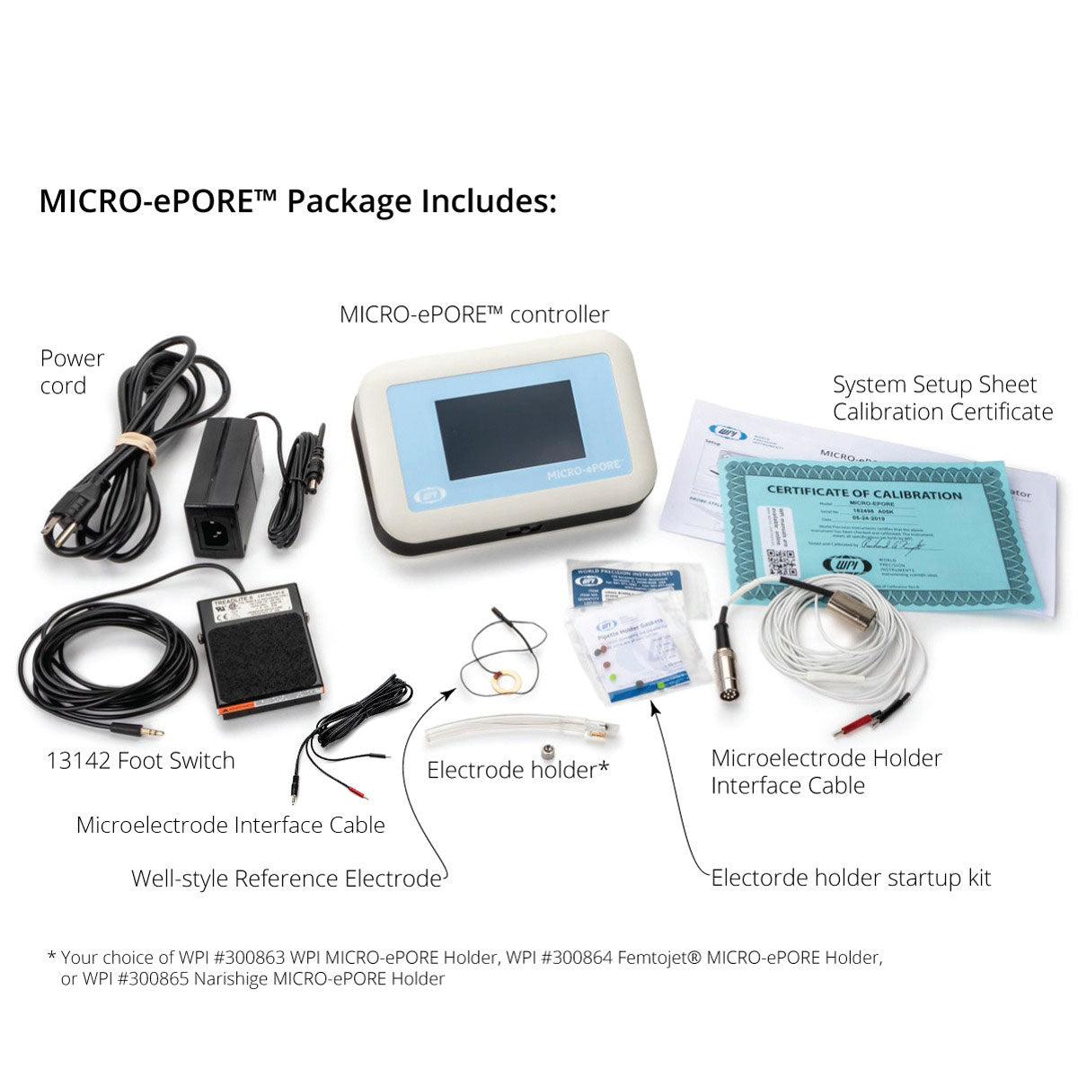
The new WPI MICRO-ePORE™ Pinpoint Cell Penetrator is a simple and versatile system that can be used for efficient microinjection of a diverse array of compounds and biomolecules into oocytes and pre-implantation stage mammalian embryos. Patent pending Flutter Electrode Technology assists in small, clean, precise membrane penetration without tearing or damaging the membrane. Here Gabe sets up the system and connects all the components.
| MICRO-ePORE™ Version 2 Rev. 2 (For units with serial numbers starting at 182508) |
MICRO-ePORE™ Version 1 Rev. 1 (For units with serial numbers up to 182508) |
| Instruction Manual v2 Quick Start Guide v2 |
Instruction Manual v1 Quick Start Guide v1 |
Is a reduction in embryo lysis possible using the MICRO-ePORE W.Gardiner, J.Kenyon, T.Bell, S.Atkins
Unpacking Your MICRO-ePORE™ System
Connecting Your MICRO-ePORE™ System
Connecting the MICRO-ePORE™ Electrode Holders
(1) MICRO-ePORE™ controller
(1) Electrode holder with silver wire. Your choice of:
• WPI #300863 WPI MICRO-ePORE Holder
• WPI #300864 Femtojet® MICRO-ePORE Holder
• WPI #300865 Narishige MICRO-ePORE™
(1) Microelectrode Holder Interface Cable
(1) Well-style Reference Electrode
(1) Probe-style Reference Electrode Assembly
(1) 13142 Foot Switch
(1) Power cord
(1) 99789 MICRO-ePORE™ Ground Cable
(1) Instruction Manual
| Specifications | Description |
| Voltage parameters | 0–3.0 V, at 1 mV increments |
| Frequency parameters | 50–3000 Hz, at 1 Hz increments |
| Pipette resistance alarm threshold maximum | 500 MΩ |
| Dimensions | 19.7 × 12.7 × 7.6 cm (7.75 × 5 × 3 in.) |
| Weight | 0.9 kg (2 lb.) |
| Certifications | CE, RoHS |
Bałakier H, Pedersen RA. Allocation of cells to inner cell mass and trophectoderm lineages in preimplantation mouse embryos. Dev Biol. 1982 Apr; 90(2):352-62. PMID: 7075865 (https://www.ncbi.nlm.nih.gov/pubmed/7075865)
Lawson KA, Pedersen RA. Cell fate, morphogenetic movement and population kinetics of embryonic endoderm at the time of germ layer formation in the mouse. Development. 1987 Nov;101(3):627-52. PMID:3502998 (https://www-ncbi-nlm-nih-gov.myaccess.library.utoronto.ca/pubmed/3502998)
Wianny F, Zernicka-Goetz M. Specific interference with gene function by double-stranded RNA in early mouse development. Nat Cell Biol. 2000 Feb; 2(2):70-5. PMID:10655585 (https://www.ncbi.nlm.nih.gov/pubmed/10655585)
Chazaud C, Yamanaka Y, Pawson T, Rossant J. Early lineage segregation between epiblast and primitive endoderm in mouse blastocysts through the Grb2-MAPK pathway. Dev Cell. 2006 May; 10(5):615- 24. PMID:16678776 (https://www.ncbi.nlm.nih.gov/pubmed/16678776)
Swann K, Campbell K, Yu Y, Saunders C, Lai FA. Use of luciferase chimaera to monitor PLCzeta expression in mouse eggs. Methods Mol Biol. 2009; 518:17-29. doi: 10.1007/978-1-59745-202-1_2. PMID:19085135 (https://www.ncbi.nlm.nih.gov/pubmed/19085135)
Posfai E, Petropoulos S, de Barros FRO, Schell JP, Jurisica I, Sandberg R, Lanner F, Rossant J. Position- and Hippo signaling-dependent plasticity during lineage segregation in the early mouse embryo. Elife. 2017 Feb 22; 6. pii: e22906. doi: 10.7554/eLife.22906. PMID: 28226240 (https://www.ncbi.nlm.nih.gov/pubmed/28226240)
Gu B, Posfai E, Rossant J. Efficient generation of targeted large insertions by microinjection into two-cell-stage mouse embryos. Nature Biotechnology 2018 Aug; 36(7):632-637. doi: 10.1038/nbt.4166. Epub 2018 Jun 11. PMID: 29889212 (https://www.ncbi.nlm.nih.gov/pubmed/29889212)

$4,600.00
Watch as Barney and Kelly Boyce set up an InVivo microscope chamber. Ideal for live cell imaging, the chambers, along with heaters, carbon dioxide and oxygen controllers and stagetop environments are sold by World Precision Instruments.
Black coated surgical instruments are not only visually more attractive than the stainless-steel ones, there are several other benefits as provided below.
Selling fast!
Get yours while you can.